Elsevier
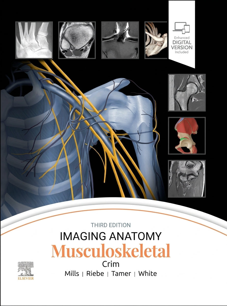
Imaging Anatomy: Musculoskeletal, 3rd Edition
Imaging Anatomy: Musculoskeletal, 3rd Edition
Quote response within 24h · Institutional invoicing available
Couldn't load pickup availability
Author: Julia Crim
ISBN: 9780443433450
Published: August 2026
Format: Hardcover, 816 pages
Language: English
Publisher: Elsevier
Description
The third edition of Imaging Anatomy: Musculoskeletal is a must-have reference for your practice. New chapters, updated content, and all-new 3T and 7T MR images provide detailed insight on both human anatomy and imaging pitfalls. Dr. Julia Crim and her team of specialists present an easy-to-read, complete anatomic atlas of the musculoskeletal system that features more than 1,900 images and illustrations as well as carefully chosen references for further reading. It is a highly visual and accessible resource for radiologists at all levels of experience as well as orthopedic surgeons, sports medicine practitioners, and all others involved with musculoskeletal imaging.
Key Features
Features all-new 3T and 7T MR images that provide exceptional imaging detail, and illustrates pediatric skeletal development. Presents radiographic, CT, and MR images complemented by large, high-quality, extensively labeled artists' illustrations throughout. Includes new chapters on the brachial plexus, scapula, clavicle, and sternum that reflect the enlarging scope of musculoskeletal practice, as well as new introductory chapters on bone maturation, bone marrow, and the musculotendinous unit. Provides detailed anatomy in at least 3 planes in MR atlas chapters, and discusses common normal variants, imaging pitfalls, and clinical applications for each area.
Coverage
Twelve sections spanning the musculoskeletal system: introduction to the musculoskeletal system, shoulder, arm, elbow, forearm, wrist, hand, pelvis and hip, thigh, knee, leg, and ankle and foot. Overview chapters for each region provide essential information, radiographs, and expert medical illustrations, alongside focused chapters diving deeper into complex anatomic issues such as the posterolateral corner of the knee.
About the Author
Dr. Julia Crim is a musculoskeletal radiologist and leads a team of specialists in presenting this comprehensive anatomic atlas, drawing on extensive experience in musculoskeletal imaging and radiology education.
Table of Contents
Section 1: Introduction to Musculoskeletal System
Section 2: Shoulder
Section 3: Arm
Section 4: Elbow
Section 5: Forearm
Section 6: Wrist
Section 7: Hand
Section 8: Pelvis and Hip
Section 9: Thigh
Section 10: Knee
Section 11: Leg
Section 12: Ankle and Foot
Why buy this book?
With more than 1,900 images including new 3T and 7T MR sequences, this atlas gives radiologists, orthopedic surgeons and sports medicine practitioners a single, highly visual reference covering every region of the musculoskeletal system, complete with the pediatric skeletal development and brachial plexus coverage that earlier editions lacked.
Keywords
Musculoskeletal radiology, imaging anatomy, MR anatomy atlas, orthopedic imaging, sports medicine imaging, bone and joint imaging, radiology atlas
Target Audience
Radiologists, orthopedic surgeons, sports medicine practitioners, musculoskeletal imaging specialists, academic medical libraries
Genre
Musculoskeletal Radiology, Orthopaedics
Q&A
Where can I buy Imaging Anatomy: Musculoskeletal, 3rd ed.?
You can order it directly from CLNZ Books, with worldwide shipping and payment by card, PayPal, or bank transfer.
What's new in the 3rd edition?
All-new 3T and 7T MR images, new chapters on the brachial plexus, scapula, clavicle and sternum, and new introductory chapters on bone maturation, bone marrow and the musculotendinous unit.
Who is this atlas for?
Radiologists at all levels of experience, orthopedic surgeons, sports medicine practitioners, and anyone involved with musculoskeletal imaging.
Does the atlas cover pediatric anatomy?
Yes, it illustrates pediatric skeletal development alongside adult anatomy throughout.
How many images does the atlas include?
More than 1,900 images and illustrations, including radiographic, CT and MR images alongside extensively labeled artists' illustrations.
📘 Learn more about shipping, delivery times, and returns, see our FAQ here